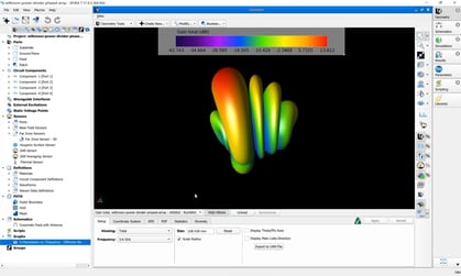
This example shows Specific Absorption Rate (SAR) simulations for a first approximation Brain Computer Implant (BCI). It was generated using an open-source and anonymized data set of patient head models that include five tissue compartments (scalp, skull, CSF, grey and white matter) [1].
Typically, human models may be represented as groups of voxels (i.e, volumetric or 3D pixels). This allows the user to distinguish tissues from one another or create detailed structures representing human anatomy. XFdtd allows import of arbitrary matrices so that users may import volumes from Python or MATLAB. Tissue segmentation of medical images such as MRI, CT, or hybrid methods often result in detailed voxel-based representations of human models [2].
Highly detailed/segmented models like the scatterBrains data are often used to train machine learning (ML) or neural network (NN) models to generate anatomically accurate human models or electromagnetic fields within human tissue [3].
 Conclusions:
XFdtd supports the import of arbitrary tissue matrices from Python or Matlab making it an excellent tool for simulating electromagnetic fields in detailed human models.
Other Details:
- Head model resolution: 1x1x1 mm^3
- Antenna is a dual resonance PIFA
- The antenna is outside the body
Sources of XFdtd-Supported Human Models:
- ICRP (free for commercial and academic)
- *Visible Human Project (free for commercial and academic)
- NICT (free for academics)
- Virtual Population (fees for both academic and commercial)
*The Visible Human Project (Male and Female) is included with the install of XFdtd and includes models at varying tissue resolutions.

References:
[1] Melissa M. Wu, Roarke W. Horstmeyer, Stefan A. Carp, "scatterBrains: an open database of human head models and companion optode locations for realistic Monte Carlo photon simulations," J. Biomed. Opt. 28(10) 100501 https://doi.org/10.1117/1.JBO.28.10.100501
[2] Lenchik, Leon et al. “Automated Segmentation of Tissues Using CT and MRI: A Systematic Review.” Academic radiology vol. 26,12 (2019): 1695-1706. doi:10.1016/j.acra.2019.07.006
[3] Di Barba, Paolo et al. “Electromagnetic Wave Absorption in the Human Head: A Virtual Sensor Based on a Deep-Learning Model.” Sensors (Basel, Switzerland) vol. 23,6 3131. 15 Mar. 2023, doi:10.3390/s23063131